lipid rich adrenal adenoma
On MR lipid-rich adrenal adenomas may demonstrate out-of-phase signal dropout which again demonstrates that the lesion is a benign adenoma despite FDG avidity Fig. Clusters of cells with enlarged lipid-rich cytoplasm seen in Cushing syndrome.

Chemical Shift Mr And Precontrast Ct Scans Of Right Lipid Rich Adrenal Download Scientific Diagram
If it does not lose SI all one can state is that the lesion does not contain lipid and is therefore not a lipid-rich adenoma.
. Fatty are the more likely it is that the tumor is not a cancer but rather the more common adrenocortical adenoma. The best treatment options for adenomas of the adrenal gland depend on many factors including whether or not the adenoma is functional releasing hormonesPeople with adrenal adenomas that are not releasing hormones usually do not require any immediate treatment. These values were consistent with lipid-rich adenoma. The approach to the radiological and clinical evaluation of adrenal masses in the oncologic and non-oncologic patient is discussed.
Lipid-rich adenomas lose signal on the chemical-shift or out-of-phase opposed-phase images while lipid-poor lesions will not lose signal. These lesions are incidentally detected in up to 44 of patients undergoing imaging studies that are performed for unrelated reasons Frequently adenomas contain abundant intracytoplasmic lipid and thus approximately 70 of adrenal adenomas are lipid-rich and are readily diagnosed because. However they may need to follow up with a healthcare provider regularly to. The front line treatment for Adrenal Adenoma is surgery.
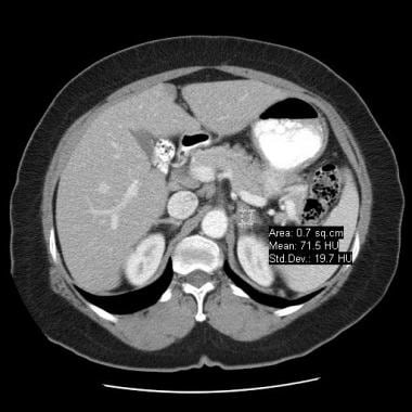
Fortunately density evaluation of an adrenal lesion is highly sensitive and specific as 70 of adrenal adenomas contain significant intracellular fat. Adrenal Adenoma is a pathological condition of the adrenal glands in which there is development of benign tumors in the adrenal glands. Adrenal metastases should not demonstrate out-of-phase signal dropout on MR. Learn what causes them how to know if you might have one and how theyre treated.
MRI has also been used to further characterise incidental adrenal masses with comparable sensitivity and specificity to unenhanced CT in identifying lipid-rich adrenal adenomas. If the testing shows an overproduction of hormones the care team may recommend removal of the affected adrenal gland although the necessity of this surgery varies. Thirty-five surgically resected adrenal adenomas were used. Though the majority are clinically.
D E Axial T1 and T2-weighted MR images showed a well-defined left adrenal mass displaying isointense signal relative to spleen on T1 and T2WIs. 10 HU lipid rich Malignancies. 96 Washout 10 15 minutes after contrast. The surgical procedure done for removal of Adrenal Adenoma is called as adrenalectomy.
Non-adenomas for instance metastases generally enhance rapidly but take longer to washout. To evaluate the relationship between lipid-rich cells of the adrenal adenoma and precontrast computed tomographic CT attenuation numbers. When an adrenal adenoma is found a series of blood urine or salivary tests are performed to assess hormone production. Adrenal metastases should not have Hounsfield units of less than 10 on unenhanced CT.
Adrenal adenomas are benign tumors of the adrenal glands which can be either functioning or non-functioning. Absolute washout A dedicated adrenal washout CT protocol consists of a non-contrast a contrast -enhanced scan with a delay of 60-90 sec and a delayed scan at 15 minutes. Thus if a lesion loses SI it is a lipid-rich adenoma Fig. Management Of The Clinically Inapparent Adrenal Mass Incidentaloma 2002 Adrenal Protocol CT Scans Initial HU without contrast.
For lipid-rich adenomas of the adrenal glands measuring under 4 cm in a patient with no underlying malignancy no follow up imaging is required 1. Lipid-poor adenomas are more difficult to diagnose as the CT density increases and approach that of soft tissue. An adrenal gland adenoma is a tumor on your adrenal gland that isnt cancer but can still cause problems. The lower the Hounsfield Units lipid-rich.
To evaluate the relationship between lipid-rich cells of the adrenal adenoma and precontrast computed tomographic CT attenuation numbers in three clinical groups. Nonenhanced CT was followed by early enhanced CT at 5 10 15 and. Sixty-five patients with 77 adrenal masses 16 lipid-poor and 37 lipid-rich adenomas and 24 nonadenomas were consecutively examined with dynamic helical CT. 18 HU Sensitivity.
Adenomas both lipid-rich and lipid-poor rapidly wash out contrast. Therefore additional endocrinological investigations are strongly recommended when an FDG-avid lipid-rich incidentaloma is detected on FDG PETCT. A lipid-rich adrenal tumor presenting increased FDG uptake compared with that of the liver is likely to be a hormone-secreting adenoma. Know the causes symptoms treatment and prognosis of adrenal adenoma.
Adenomas are the most common tumors arising from the adrenal cortical tissue. In comparison to surrounding adrenal gland adenoma cells are larger with different cytoplasm increased variation in nuclear size Distinct cell borders cells have abundant foamy cytoplasm reminiscent of zona fasciculata Balloon cells. In addition the value of unenhanced and enhanced CT densitometry with emphasis on the washout features to distinguish between lipid-rich and lipid-poor adenomas and malignant lesions is detailed. Up to 10 cash back The T2W properties of a lipid-rich adenoma may be useful to establish diagnosis when differentiating lipid-rich adenoma from other adrenal masses which may demonstrate microscopic fat on chemical-shift MRI or when chemical-shift MRI is degraded by artifact or limited by through-plane resolution in small adrenal nodules.
For lipid-poor lesions the contrast washout rate can be calculated using CT. 53 rows If an adrenal mass measures 10 HU or less on unenhanced CT it is probably a lipid-rich adenoma14 The mean Hounsfield unit for adrenal carcinoma metastasis and pheochromocytoma is significantly. A high-quality CT scan using contrast is the most important x-ray or scan. 15 MRI is not as effective as CT washout in characterising lipid-poor adrenal lesions and for incidental lesions measuring 30 Hounsfield units CT adrenal protocol.
The roles of magnetic. The clinical diagnoses of the patients included 13 cases of primary aldosteronism 15. The marked reduction in signal intensity between the in-phase and out of phase T1-weighted images indicates fatty content and therefore a lipid-rich adenoma. By quantitative assessment of chemical shift images the adrenal-to-spleen ratio ASR was 031 and signal intensity index SII was 42.
In cases when a patient with an adrenal adenoma also is.

Adrenal Incidentaloma Adrenal Tumor Found By Accident

X Rays Ct Scans Mri And Other Tests For Adrenal Glands

Adrenal Adenoma Radiology Case Radiopaedia Org
2

Adrenal Adenoma Imaging

Posting Komentar untuk "lipid rich adrenal adenoma"